Anatomy Of Chest Cavity / Thoracic Cavity at Salt Lake Community College - StudyBlue - Thoracic cavity, the second largest hollow space of the body.
Anatomy Of Chest Cavity / Thoracic Cavity at Salt Lake Community College - StudyBlue - Thoracic cavity, the second largest hollow space of the body.. Dysfunctional breathing an online course for physical therapists / physiotherapists powered by physiopedia start. If you have pain in the chest, see your doctor or. A good radiologist knows the anatomy, so don't skip this chapter! They are located in the chest, either side of the mediastinum. However, what is the anatomic definition or meaning of a 'chest'?
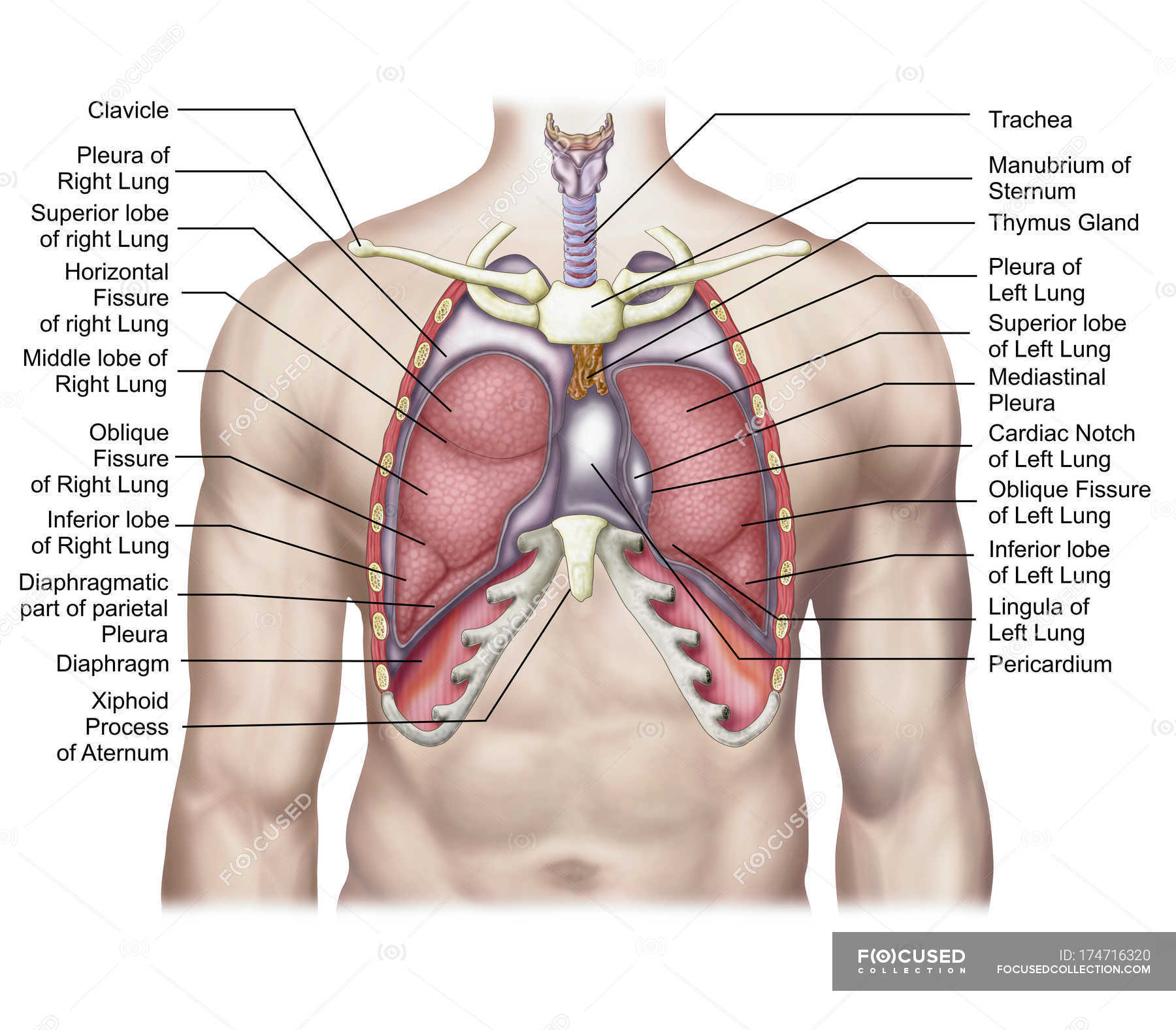
A good radiologist knows the anatomy, so don't skip this chapter! Advanced anatomy & physiology tony serino, ph.d. 'heart disease anatomical chart 2nd edition poster print' posters | allposters.com. Each of these anatomical structures should be viewed using a systematic approach. It is enclosed by the ribs, the vertebral column, and the sternum, or breastbone, and is separated from the abdominal cavity by the diaphragm.
Medical illustration of human lungs anatomy with labels ... from st.focusedcollection.com ¼ to 1/3 of thoracic cavity apex to left cardiac axis. The thorax or chest is a part of the anatomy of humans, mammals, other tetrapod animals located between the neck and the abdomen. If you have pain in the chest, see your doctor or. Among the major organs contained in the thoracic cavity are the heart and lungs. The epidermis is the outermost layer that provides a protective, waterproof seal over the body. 'heart disease anatomical chart 2nd edition poster print' posters | allposters.com. This anatomical midline can be useful in assessing for symmetry in breast augmentation or in performing a median sternotomy. The function of the lungs is to the lungs lie either side of the mediastinum, within the thoracic cavity.
A brief tour of embryonic development of anatomical structures and organs.
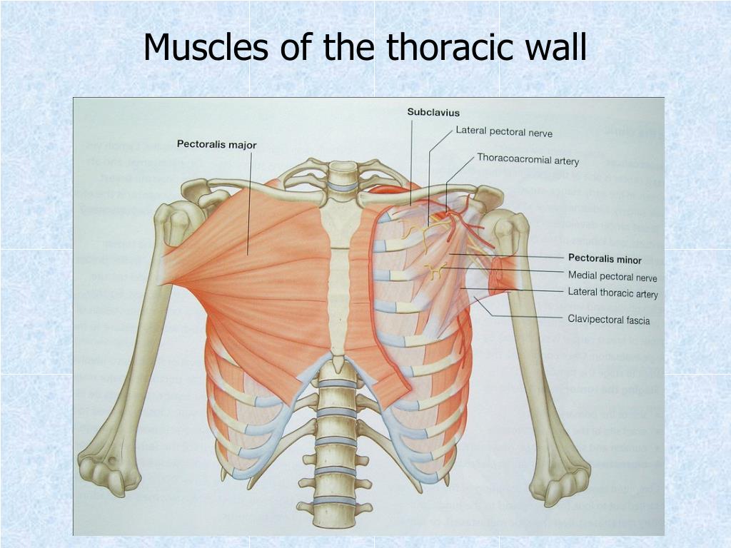
The upper ventral, thoracic, or chest cavity contains the heart, lungs, trachea, esophagus, large blood vessels, and nerves. The treatment for chest pain depends upon the cause. 'heart disease anatomical chart 2nd edition poster print' posters | allposters.com. Related online courses on physioplus. Anatomy of the peritoneal cavity. Thoracic cavity, the second largest hollow space of the body. It is enclosed by the ribs the vertebral column and the sternum or breastbone and is separated from the abdominal cavity the bodys thoracic cavity definition organs of chest cavity the chest is the area of origin for many of the bodys systems as it houses organs such as the. The vital structures of the thoracic cavity (chest cavity) can be identified at certain key points within the chest(8). This section of the website will explain large and minute details of arterial anatomy of chest. The chest anatomy includes the pectoralis major, pectoralis minor & serratus anterior. If you need to learn about the body cavities such as the thoracic cavity, also called the chest cavity, sits superior (higher) to the abdominopelvic cavity, and it contains organs such as the heart, lungs. Since there are so many of them, the thoracic cavity is divided. A brief tour of embryonic development of anatomical structures and organs.
Advanced anatomy & physiology tony serino, ph.d. Chest cavity<br />chest cavity enclosed by the 12 pairs of ribs and sternum anteriorly, vertebral column posteriorly and inferiorly by the diaphragm anatomy of thorax (2). A brief tour of embryonic development of anatomical structures and organs. Roof is called the tegmen and separates the upper part of the tympanic cavity or epitympanum from the middle cranial fossa. Related online courses on physioplus.
Pleurisy - Causes, Symptoms, Pain, Diagnosis & Treatment from healthjade.com Because the left lung does not contact the anterior portion of the left thoracic cavity at this level, the heart with its epicardial fat occupies this space. It is enclosed by the ribs, the vertebral column, and the sternum, or breastbone, and is separated from the abdominal cavity by the diaphragm. Anatomy of the chest cavity. Among the major organs contained in the thoracic cavity are the heart and lungs. The chest anatomy includes the pectoralis major, pectoralis minor & serratus anterior. Roof is called the tegmen and separates the upper part of the tympanic cavity or epitympanum from the middle cranial fossa. Each of these anatomical structures should be viewed using a systematic approach. The chest wall is formed from the sternum anteriorly, 12 pairs of ribs, costal cartilages and intercostal muscles laterally, and the thoracic vertebrae posteriorly.
Learn about each muscle, their locations & functional anatomy.
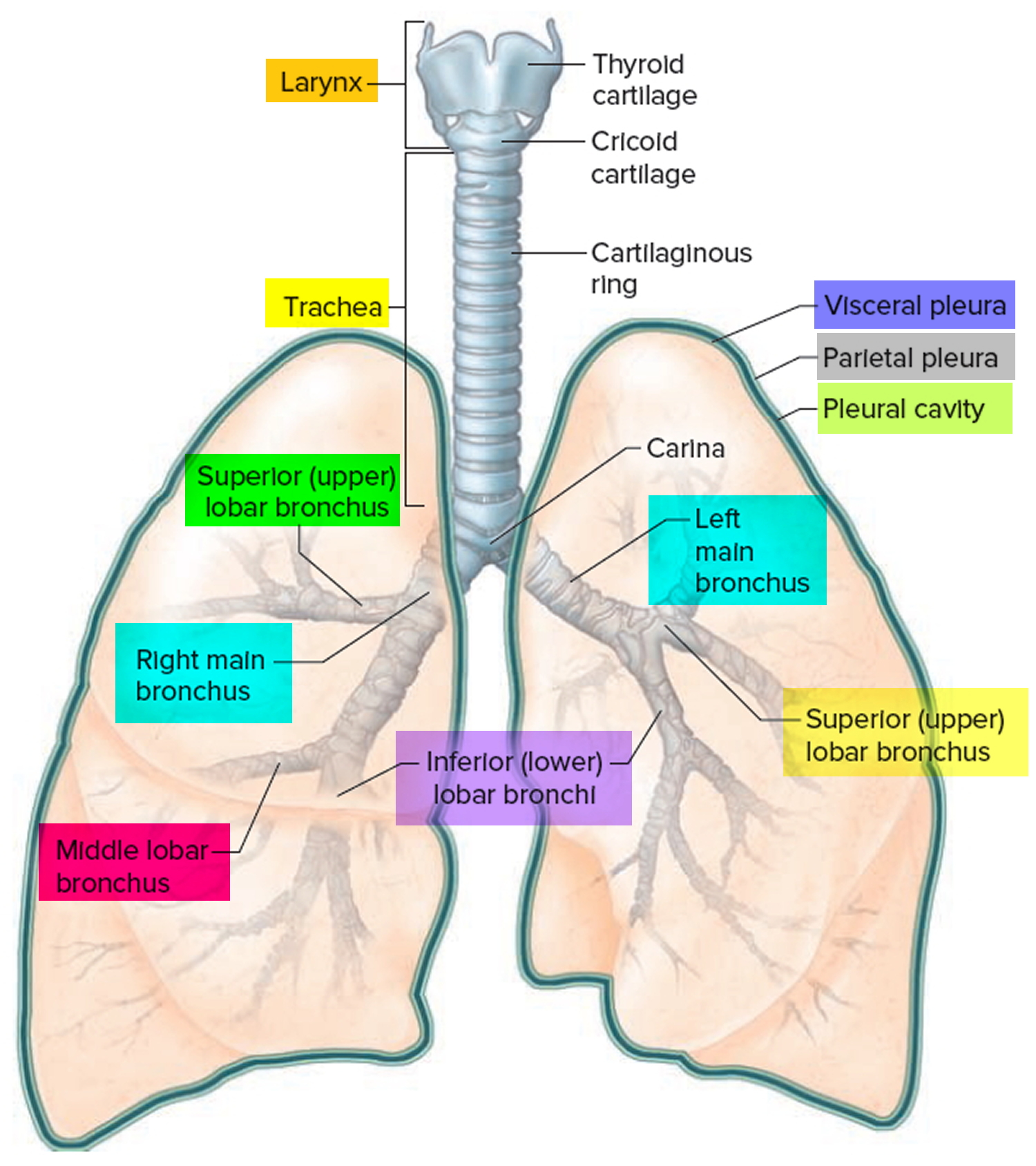
Dysfunctional breathing an online course for physical therapists / physiotherapists powered by physiopedia start. If you need to learn about the body cavities such as the thoracic cavity, also called the chest cavity, sits superior (higher) to the abdominopelvic cavity, and it contains organs such as the heart, lungs. The function of the lungs is to the lungs lie either side of the mediastinum, within the thoracic cavity. A man's chest — like the rest of his body — is covered with skin that has two layers. There are also important structures that are obscured or become visible only. The upper ventral, thoracic, or chest cavity contains the heart, lungs, trachea, esophagus, large blood vessels, and nerves. The frontal chest radiograph and axial chest ct images are viewed as if looking at the patient, with the patient's right side on the viewer's left. Chest pain can be caused by many diseases and condition, for example,angina, heart attack, shingles, pneumonia, pulmonary embolism, pericarditis, gerd, broken or bruised ribs, and aortic dissection. The vital structures of the thoracic cavity (chest cavity) can be identified at certain key points within the chest(8). Because the left lung does not contact the anterior portion of the left thoracic cavity at this level, the heart with its epicardial fat occupies this space. The epidermis is the outermost layer that provides a protective, waterproof seal over the body. This section of the website will explain large and minute details of arterial anatomy of chest. Radiology basics of chest ct anatomy with annotated coronal images and scrollable axial images to help medical students and junior doctors learning anatomy.
Anatomy of the chest cavity. Reading of chest radiographs, some basic anatomy and physiology including, pleural fissures, mediastinal lines, the bronchi and in reading chest radiographs it is important to understand their limitations, basic anatomy and. Your poster is printed with an offset lithography press with a coating to protect the inks. Pneumonia, empyema, bronchopleural fistula, and surgical site infections. A brief tour of embryonic development of anatomical structures and organs.
PPT - Chest wall, thoracic cavity and pleura PowerPoint ... from image2.slideserve.com Reading of chest radiographs, some basic anatomy and physiology including, pleural fissures, mediastinal lines, the bronchi and in reading chest radiographs it is important to understand their limitations, basic anatomy and. This anatomical midline can be useful in assessing for symmetry in breast augmentation or in performing a median sternotomy. Because the left lung does not contact the anterior portion of the left thoracic cavity at this level, the heart with its epicardial fat occupies this space. If you have pain in the chest, see your doctor or. Anatomy of the peritoneal cavity. They are located in the chest, either side of the mediastinum. It is enclosed by the ribs, the vertebral column, and the sternum, or breastbone, and is separated from the abdominal cavity by the diaphragm. Thoracic cavity, the second largest hollow space of the body.
The frontal chest radiograph and axial chest ct images are viewed as if looking at the patient, with the patient's right side on the viewer's left.
It is enclosed by the ribs the vertebral column and the sternum or breastbone and is separated from the abdominal cavity the bodys thoracic cavity definition organs of chest cavity the chest is the area of origin for many of the bodys systems as it houses organs such as the. Anatomy of the chest cavity. Learn about each muscle, their locations & functional anatomy. Roof is called the tegmen and separates the upper part of the tympanic cavity or epitympanum from the middle cranial fossa. Advanced anatomy & physiology tony serino, ph.d. Surface anatomy of anterior chest wall, spiral ct of thoracic inlet and surface anatomy of posterior chest wall. The function of the lungs is to the lungs lie either side of the mediastinum, within the thoracic cavity. A brief tour of embryonic development of anatomical structures and organs. Each of these anatomical structures should be viewed using a systematic approach. Because the left lung does not contact the anterior portion of the left thoracic cavity at this level, the heart with its epicardial fat occupies this space. The vital structures of the thoracic cavity (chest cavity) can be identified at certain key points within the chest(8). It is enclosed by the ribs, the vertebral column, and the sternum, or breastbone, and is separated from the abdominal cavity by the diaphragm. Some physiology, and to have a systematic system.
'heart disease anatomical chart 2nd edition poster print' posters | allposterscom anatomy of chest. Anatomy of the peritoneal cavity.